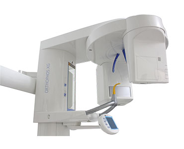
With a perfectly designed 3D cylinder volume of 8 cm in diameter and 8 cm in height and a standard resolution of 160 µm, ORTHOPHOS XG 3D is precisely tailored to the everyday routines of private practices: it can capture the patient's whole jaw in a single span. The field of view is large enough to avoid the stitching of several 3D x-ray images and thus multiple exposures to radiation. Yet it is also small enough to be a time-saver in diagnosis.
If an even smaller volume is sufficient, then choose the 5 x 5.5cm VOL 2 with a resolution of 100 µm. whenever you need to see more, the 3D module provides for greater safety. In all standard cases, the 2D module of ORTHOPHOS XG 3D, with extensive panoramic and cephalometric x-ray programs, delivers the x-ray image you need.
Striking image quality with ASTRA (2D), MARS (3D) and High-Definition mode.
Two scan volumes for dose reduction and time-efficient diagnostics.
High resolution (160/100 µm).
Simple unit operation for 2D and 3D scans with automatic switching sensor.
Automatic patient positioning for correct panoramic scans.
E.g., displaced, impacted teeth, cephalometric analysis, root resorptions, and cleft lips, jaws and palates.
General dentistry
E.g, contradictory findings, as well as those that are difficult or impossible to view in the OPG, apical lucency, periodontal diseases, patient consultations, endodontics, implantology, minor oral interventions.
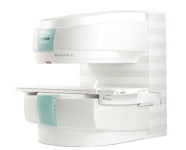
Open MRI Siemens
MAGNETOM C!™ increases confidence in diagnosis due to excellent image quality at 0.35T. With the most open, compact C-shaped magnet, it provides outstanding patient comfort. All of this comes with comparatively low power consumption and operating costs.
Confidence in diagnosis due to superb image quality at 0.35T
From now on, high-field diagnostic confidence at mid-field can be your standard with MAGNETOM C!.
Cost efficient quality care
Comprehensive workflow from start to finish. With MAGNETOM C! Every step is optimized to streamline your examinations: beginning with quick and easy patient positioning and posterior coil parts that remain on the table.
Outstanding patient comfort with the most open, compact C-shaped magnet
More space means less anxiety. A unique C-shaped magnet for easy side loading and a friendly, compact design cooperate to create an open and free atmosphere.
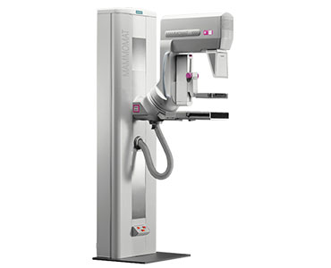
Excellent contrast. High resolution. Brilliant images.
A dedicated mammography X-ray tube with molybdenum anode and optimized imaging parameters ensure optimal beam quality. This provides images with excellent contrast and high resolution at short exposure times. The Opdose® function auto-selects the best kV value and anode/filter combination (Mo/Mo and Mo/Rh) according to the individual breast characteristics in order to achieve optimal image quality with the lowest possible dose.
Automatic Sensor :
MAMMOMAT® 1000 is equipped with OpComp, which automatically and individually senses the breast compression needed to produce the ideal combination of minimal patient discomfort and outstanding image quality.
Compression plates :
A wide array of compression plates ensures gentle compression optimized for each individual breast. An optionally available perforated biopsy compression plate and magnification tables further expand the scope of applications.
Excellent access :
The open design means excellent access, even for patients in a wheelchair. Examinations can be performed with equal ease on patients in standing, sitting or recumbent positions.
For more details please click here and search for your desired test
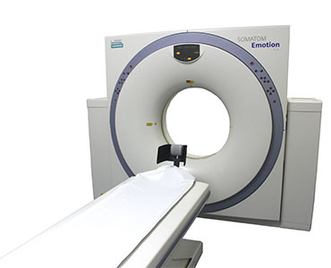
CT Scan
It is famous for its great versatility and high performance from exam set-up to image acquisition and processing – and will give you high efficiency in delivering high quality clinical outcomes.
Innovative dose-saving features such as IRIS, CARE Dose4D and DoseMAP, combine the best possible image quality at the lowest possible dose. Simply fabulous.
FAST. FAMOUS. FABULOUS.
FAST – high efficiency and high quality care
Committed to continuous innovation, SOMATOM® Emotion now runs with the award winning FAST CARE1 technology, providing new features such as FAST Planning and FAST Spine.
Famous – great reputation and great value
From the imaging market leader, SOMATOM Emotion is installed at at nearly 9,000 institutes around the world - and it is running and running. Famous due to its great versatility and high performance, it is both a success story and great value for money.
Fabulous – leading image quality and leading dose technology
With SOMATOM Emotion, you get leading image quality. It delivers a very small focal spot and thus outstanding routine spatial resolution.
Here are the parts of the body where you can use our CT SCAN. For details please click here and search for your desired test.
• BOTH KNEES
• ABDOMEN and PELVIS
• BRAIN
• CERVICAL VERT
• CHEST
• COCCYX VERT
• CONTRAST PROCEDURE CT
• DENTAL
• HIP
• ANY PERIPHERAL PART (HAND/WRIST)
• JOINTS (ANKLE, KNEE, SHOULDER)
• LARYNX
• LUMBAR VERT
• NASOPHARYNX
• ORBIT
• OSTEO
• PELVIS
• PETROUS BONE & ORBIT & SELLA
• PETROUS BONE (EAR)
• P.N.S. CORONAL & AXIAL
• SCANOGRAM
• SELLA (SKULL)
• THORACIC VERT
• VERTEBRA (Two Parts)
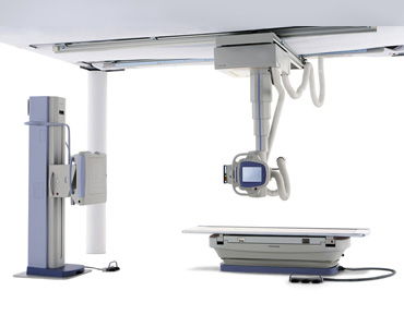
Digital X-Ray Images
X-ray imaging provides fast, high-resolution images and is relatively inexpensive. The average examination for most plain film examinations takes no more than 10–15 minutes and requires no special preparation of the patient. The operator, usually the radiographer (also known as a radiologic technologist), selects the amount and type of X-rays to be used according to the patient’s size, the tissue or part of the body being imaged and the amount of image contrast required. Because movement, e.g. of the lungs and diaphragm, blurs the image, patients are usually asked to hold their breath during the exposure. The X-ray picture is stored on a piece of film called a radiograph. These are interpreted by a physician specially trained to interpret them, known as a radiologist.
For more details please click here and search for your desired test
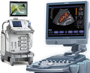
Ultrasonography
Ultrasound is safe and painless, and produces pictures of the inside of the body using sound waves. Ultrasound imaging, also called ultrasound scanning or sonography, involves the use of a small transducer (probe) and ultrasound gel placed directly on the skin. High-frequency sound waves are transmitted from the probe through the gel into the body. The transducer collects the sounds that bounce back and a computer then uses those sound waves to create an image.
Ultrasound imaging has been used for over 20 years and has an excellent safety record. It is based on non-ionizing radiation, so it does not have the same risks as X-rays or other types of imaging systems that use ionizing radiation.
Ultrasound imaging is a medical tool that can help a physician evaluate, diagnose and treat medical conditions. Common ultrasound imaging procedures include:
Abdominal ultrasound (to visualize abdominal tissues and organs).
Bone sonometry (to assess bone fragility).
Breast ultrasound (to visualize breast tissue).
Doppler fetal heart rate monitors (to listen to the fetal heart beat).
Doppler ultrasound (to visualize blood flow through a blood vessel, organs, or other structures).
Echocardiogram (to view the heart).
Fetal ultrasound (to view the fetus in pregnancy).